
Equipment List
- HOME
- Equipment List

MOVIE
Moonshot Webinar “Introduction of Shared Equipment at Kento Imaging Support Center”
(Time : 1:00:30)
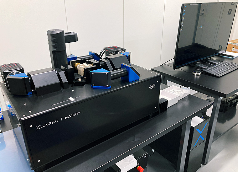
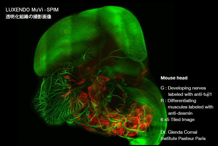
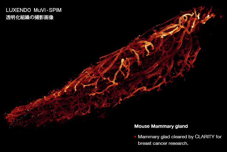
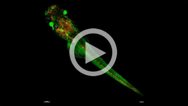
Multi View Light Sheet Microscope
MuVi SPIM LS&CS
Luxendo
MuVi SPIM LS&CS light sheet microscope from Luxendo, a Bruker company, enables high-resolution imaging of live small animals (e.g. zebrafish) and mouse brains and organs after tissue clearing. This microscope is equipped with a motorized XYZ stage that can rotate 360° to accommodate the mounting devices, 405, 488, 561, and 642 nm excitation lasers, and a high-speed filter wheel for up to 10 colors.
| Laser (nm) | Filter (nm) | Detector | Objective lens |
|---|---|---|---|
| 405 (405-50) 488 (488-60) 561 (561-60) 642 (642-70) |
BP 418-462 LP 498 BP 497-554 LP 572 BP 580-627 LP 656 BP 655-704 |
ORCA-flash4.0 V3 | 10x, ND 0.50, WD 5.5 mm 20x, ND 1.00, WD 8.2 mm |

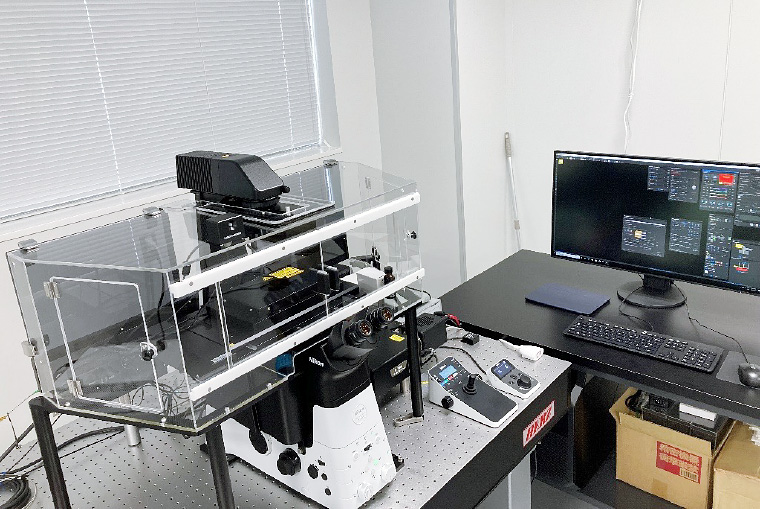
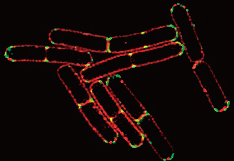
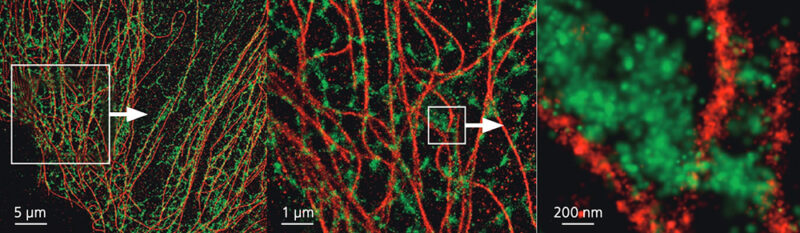
super-resolution microscope
N-SIM S/N-STORM
Nikon Corporation
Nikon's super-resolution microscopes [N-SIM S/ N-STORM] integrate Structured Illumination Microscopy (SIM), which enables high-speed image acquisition at up to 15 fps, and Stochastic Optical Reconstruction Microscopy (STORM), a high-resolution microscopy technique with horizontal resolution of 20 nm and Z-axis resolution of 50 nm, into a single system for various super-resolution imaging applications. Stochastic Optical Reconstruction Microscopy (STORM) is integrated into a single system for a variety of super-resolution imaging applications. An automatic focus maintainer that precisely compensates for the effects of temperature changes and vibration, and an objective lens with a motorized correction ring that corrects spherical aberration with a single click, support nano-scale imaging.
| Laser (nm) | Filter | Detector | Objective lens |
|---|---|---|---|
| [SIM] 405 488 561 647 [STORM] 405 488 561 647 |
Dapi / GFP / RFP / Cy5 | ORCA-FusionBT | Plan Apo 10x, NA 0.45, WD 4.00 S Plan Fluor ELWD 20x Ph1 Adm (air), NA 0.45, WD 8.20 SR HP Plan Apo Lambda S 100xC (silicone oil), NA 1.35, WD 0.30 SR HP Apo TIRF 100xAC (oil), NA 1.49, WD 0.12 |
- Features (link to manufacturer’s website)
・N-SIM S/N-STORM - Catalog (English) PDF
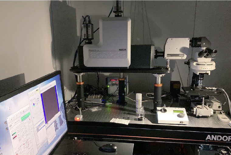
High-speed confocal scanning microscope
Dragonfly
Andor Technology Ltd.
Dragonfly is a spinning disk confocal microscope system capable of high-speed imaging up to 400 fps. The system is equipped with four types of lasers (405 nm, 445 nm, 488 nm, and 561 nm) and a high-resolution, high-dynamic-range sCMOS camera on a fixed-stage upright microscope base, enabling low phototoxicity and high contrast high-speed 3D imaging of a wide variety of samples, from intracellular dynamic observation to tissue- and organ-level observation.
| Mocroscope | Laser (nm) | Filter (nm) | Detector | Objective lens |
|---|---|---|---|---|
| Dragonfly (Andor) | 405nm / 445nm / 488nm / 561nm | BFP / CFP / GFP / RFP | sCMOS camera/ ZYLA4.2P-USB3 | Nikon LWD 16x (water), NA 0.80, WD 3 mm Nikon Apo LDW 25x (water), NA 1.10, WD 2 mm |
Raman microscope
Nikon Corporation
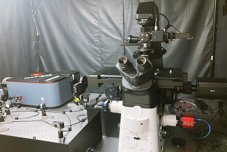
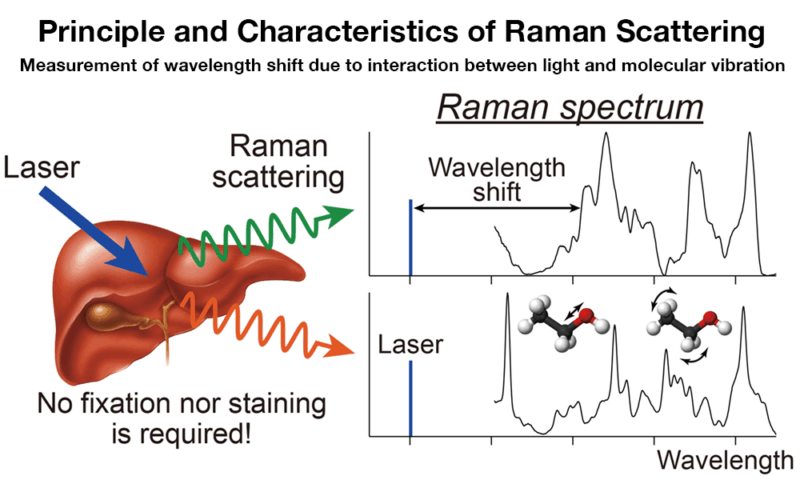
The Raman microscope is an optical microscope that observes Raman spectra reflecting molecular information (molecular vibrations) of the sample. Samples can be fixed or unfixed, cells, tissue sections, excised tissues, etc., as long as they can be placed on the inverted microscope. Our Raman microscope uses a 532 nm laser for excitation (other wavelengths will be added in the future). Fluorescence imaging using an LED light source and multiphoton observation using a 785 nm femtosecond laser can also be performed at the same time.


electron microscope
JSM-IT800
JEOL Ltd.
JSM-IT800 is a JEOL's latest high-performance scanning electron microscope, which enables not only conventional observation of surface 3D structures but also acquisition of images similar to section images obtained by transmission electron microscopy over a larger area when ultra-thin sections are used as samples, and is equipped with an option for the serial section imaging. For the time being, we plan to offer this service on a contract basis.
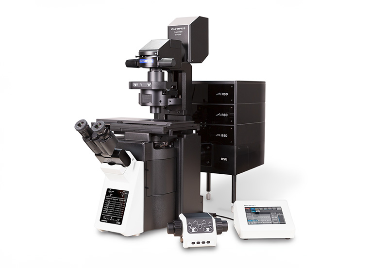
multiphoton excitation microscope
FVMPE-RS-SS-SP
Olympus(Evident Corporation)
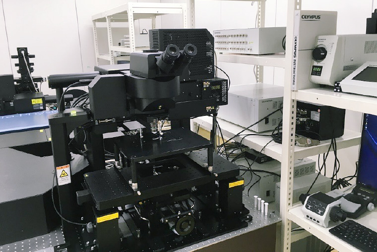
The Olympus multiphoton excitation microscope FVMPE-RS-SS-SP is equipped with an InSight X3 laser covering a wide excitation wavelength range from 680 nm to 1300 nm and four GaAsP detectors with high optical sensitivity, enabling high-resolution and multicolor 3D fluorescence imaging. Both upright and inverted microscopes and two types of scanning units (high-precision galvanometer scanner + high-speed resonant scanner) are available for various experimental protocols.
| Laser (nm) | Filter | Detector |
|---|---|---|
| Main (680-1300) Sub (1045) |
CFP/YFP/RFP/SHG GFP/RFP/SHG etc. (Consultation requested) |
GaAsP Photomultipliyer 4ch (GaAsP PMT) |
| Objective lens |
|---|
| [Upright] XLPLN 10XSVMP (water, silicon oil, oil), NA 0.60, WD 8.00 XLPLN 25XWMP2 (water), NA 1.05, WD 2.00 [Inverted] UPLXAPO 10X (air), NA 0.40, WD 3.10 UPLSAPO 30XS (silicone oil), NA 1.05, WD 0.80 |
Confocal Laser Scanning Microscope
FV3000
Olympus(Evident Corporation)
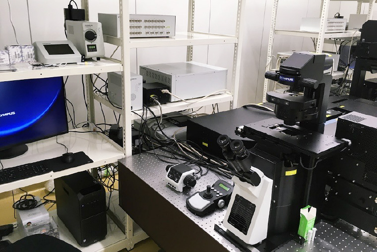
FV3000 confocal laser scanning microscope is capable of extremely sensitive and highly accurate confocal imaging, enabling acquisition of weak fluorescence signals and imaging with lower laser power to reduce damage to a specimen and fading of fluorescence. The system is also equipped with a precise stitching function using multi-area imaging, making it suitable for wide range imaging from the cellular level to large tissues. The intuitive, easy-to-use software allows for a variety of imaging options, including Z-stack, time-lapse, and stitching.
| Mocroscope | Laser (nm) | Filter (nm) | Detector |
|---|---|---|---|
| FV3000 (EVIDENT) | 405 nm 488 nm 561 nm 640 nm 445 nm 514 nm 594 nm |
CFP / YFP / GFP / RFP / Cy5 etc. (Consultation requested) |
GaAsP Photomultipliyer 4ch (GaAsP PMT) |
| Objective lens |
|---|
| UPLXAPO 10X (air), NA 0.40, WD 3.10 UPLXAPO 20X (air), NA 0.80, WD 0.60 UPLXAPO 40X (air), NA 0.95, WD 0.18 UPLSAPO 30XS (silicone oil), NA 1.05, WD 0.80 UPLSAPO 60XS2 (silicone oil), NA 1.30, WD 0.30 PLAPON 60XOSC2 (oil), NA 1.40, WD 0.12 UPLXAPO100XO (oil), NA 1.45, WD 0.17 UPLXAPO40XO (oil), NA 1.40, WD 0.17 UPLSAPO100XS (silicone oil), NA 1.35, WD 0.13-0.19 PLAPON 2X (air), NA 0.08, WD - UPLXAPO 4X (air), NA 0.16, WD - UCPLFLN 20X (air), NA 0.70, WD 0-1.6 |
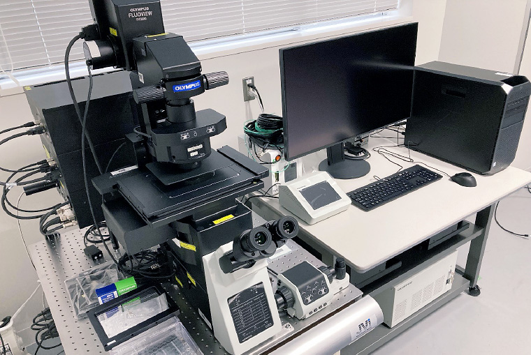
Confocal Laser Scanning Microscope
FV4000
Olympus(Evident Corporation)
FV4000 confocal laser microscope is equipped with a semiconductor sensor SilVIR detector, which enables extremely low-noise, high-sensitivity confocal imaging and quantification of images by photon counting. This enables high-speed imaging at high image quality even with a 1K-resonant scanner. The wide dynamic range enables multi-scale imaging from the organ/tissue level to the intracellular microstructure level. The FV4000 is a great choice for more quantitative and reproducible imaging.
| Mocroscope | Laser (nm) | Filter (nm) | Detector |
|---|---|---|---|
| FV4000 (EVIDENT) |
405 nm 488 nm 561 nm 640 nm 445 nm |
CFP / YFP / GFP / RFP / Cy5 etc. (Consultation requested) |
SilVIR detector |
| Objective lens |
|---|
| UPLSAPO 10X (air), NA 0.40, WD - UPLSAPO 20X (air), NA 0.75, WD 0.60 UPLSAPO 40X (air), NA 0.95, WD 0.18 UPLSAPO 30XS (silicone oil), NA 1.05, WD 0.80 UPLSAPO 60XS2 (silicone oil), NA 1.30, WD 0.30 UPLXAPO 60XO (oil), NA 1.42, WD 0.15 UPLXAPO100XO (oil), NA 1.45, WD 0.17 UPLXAPO40XO (oil), NA 1.40, WD 0.17 UPLSAPO100XS (silicone oil), NA 1.35, WD 0.13-0.19 PLAPON 2X (air), NA 0.08, WD - UPLXAPO 4X (air), NA 0.16, WD - |